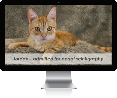

Transcolonic portal scintigraphy
is a noninvasive technique for diagnosing portosystemic shunts in dogs and cats that does not require anesthesia or sedation.
What is a portosystemic shunt and how would it affect my animal?
A portosystemic shunt is an abnormal communication between the portal circulation and the systemic circulation (the blood supply to the rest of the body). This communication is usually an abnormal blood vessel that is present at birth (congenital). Occasionally the abnormal blood vessel may develop during the life of the animal (acquired) as a result of an ongoing liver disease. In either case the shunt vessel allows the blood from the gastro-intestinal tract to bypass the liver and go directly into the systemic circulation.
When a portosystemic shunt exists not only is the liver malnourished due to a lack of needed nutrients, but the rest of the body is subjected to potentially high levels of toxins that would normally have been filtered out or inactivated by the liver. Portosystemic shunts may develop in a blood vessel outside of the liver (extrahepatic) or in a blood vessel within the liver itself (intrahepatic).
The clinical signs in dogs and cats with portosystemic shunts are highly variable but usually involve poor growth and central nervous system signs that result from the presence of high levels of toxins in the blood stream. These central nervous system signs may include intermittent disorientation, pacing, circling, tremors and even seizures. While the possibility of a portosystemic shunt can be diagnosed with routinely available laboratory blood tests, confirmation of the shunt requires further imaging techniques.
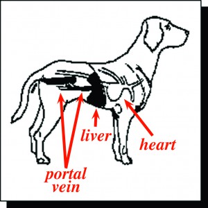
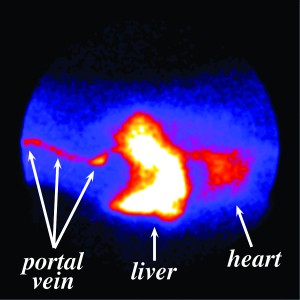
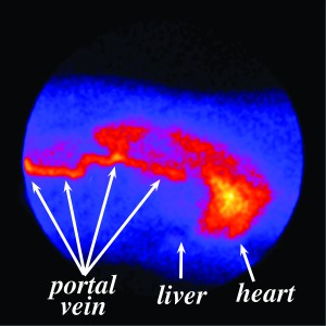
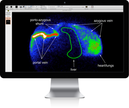
Transcolonic portal scintigraphy involves placement of a radioactive "dye" into the intestine (rectum) of an animal and recording the route of dye absorption. In a normal animal the dye is absorbed into the portal circulation and rapidly transported to the liver. In an animal with a portosystemic shunt, the dye bypasses the liver and appears in the heart first. This procedure is performed without sedation or anesthesia and takes only a few minutes to perform. Transcolonic portal scintigraphy is currently the most accurate and yet safest way available to confirm the diagnosis of a portosystemic shunt.
What can be done if my animal has a portosystemic shunt?
In cases of an extrahepatic shunt, the condition can usually be successfully treated by surgically closing the shunt vessel. This forces the blood collected from the gastrointestinal tract to flow into the liver and returns the portal blood flow to normal. In cases of an intrahepatic shunt, the surgery is significantly more complicated. Because the shunt vessel is less accessible, other procedures are usually performed to increase the portal blood flow to the liver. Most animals who have surgery performed early in the course of the disease will do well.
Are there treatment options other than surgery?
Most animals with portosystemic shunts can clinically improve with an adjustment in their diet and the administration of certain antibiotics. Unfortunately these methods do not repair the underlying disorder and usually become less effective in managing the condition over time.
If my veterinarian thinks my pet may have a portosystemic shunt, how do I arrange to get transcolonic portal scintigraphy performed?
Either you or your veterinarian can call Advanced Veterinary Medical Imaging and arrange to have this procedure performed with less than 24 hours advance scheduling.