Bone Scintigraphy (or bone scanning) is a valuable tool in the evaluation of the skeletal system. Bone scintigraphy depends on the principal that actively metabolizing bone will incorporate certain bone "tracers." The distribution of these tracers is dependent on the rate of bone turnover and blood flow. The images obtained reveal the relative distribution of these tracers throughout the skeletal system. Since bone scintigraphy reveals changes in bone metabolism more than changes in bone structure, bone scintigraphy complements rather than replaces plain radiography (X-rays). The changes noted on bone scintigraphy usually precede the changes noted on radiographs because the bone metabolism usually changes before the bone structure changes.
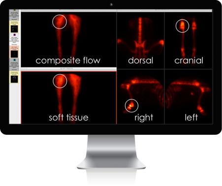
The process of bone scintigraphy involves the acquisition of images at various times after the administration of the tracer. The tracer usually requires several hours to be incorporated into the bone and cleared from the remaining tissues of the body. By evaluating the images taken at different times after the administration of the tracer, information about blood flow verses soft tissue (muscle, ligament, tendon) and bone metabolism can be obtained.
Any condition which results in a change in the metabolism of the bone will result in a change in the appearance of the bone scan. As a result lesions like fractures, infections, tumors, and arthritis can be recognized with a bone scan long before they can be seen with plain radiographs.
One of the most useful applications for bone scintigraphy is in the evaluation of the patient with a poorly localized lameness. Localization of the source of a lameness in a veterinary patient is frequently difficult. This difficulty is partially due to the inability of our patients to communicate the source of their pain and is complicated by the fact that many of our patients are extremely stoic and fail to react even when the painful area is directly manipulated. Radiographs (X-rays) are usually very helpful in localizing the cause of the lameness.
Unfortunately many common causes for lameness in dogs and cats do not result in readily demonstrable radiographic lesions until the condition is advanced. This occurs because the structural or anatomic lesion may be extremely small. Fortunately many of these lesions result in a significant change in the metabolism of the bone. This change in the metabolism is what bone scintigraphy is able to detect
By allowing the detection of the lesion early in the course of the disease, bone scintigraphy may allow for a surgical correction of the problem. Tumors are obviously best treated early in the course of the disease, before they have spread. Conditions causing lameness are usually best treated early in their course, before the development of secondary arthritis which may be incurable.