Cerebellar Hypoplasia

Etiology

Cerebellar hypoplasia is a congenital condition that is more common in cats than in dogs. Several types of malformations have been described in cats and dogs, including the following:

1) Agenesis: Part of or the entire cerebellum is absent.
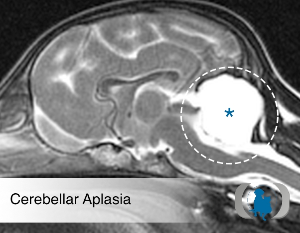
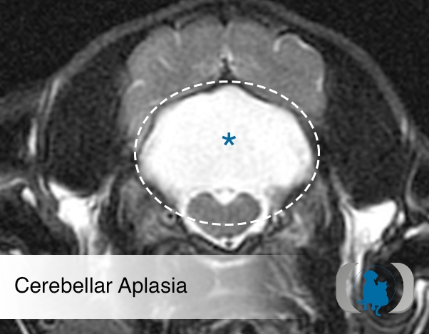
2) Aplasia: Development of cerebellum is faulty, with no tissue differentiation.
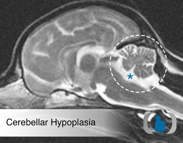
3) Hypoplasia: Faulty development is present but some tissue differentiation has occurred. The cerebellum is smaller than normal.
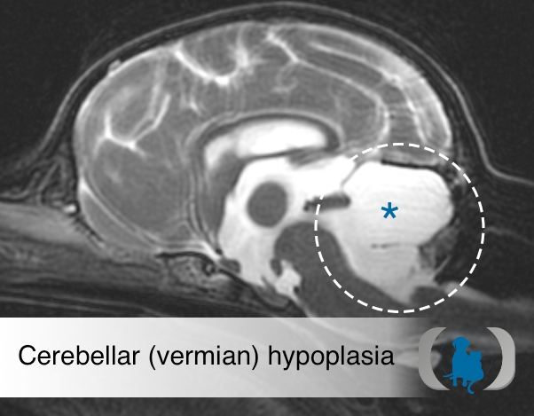
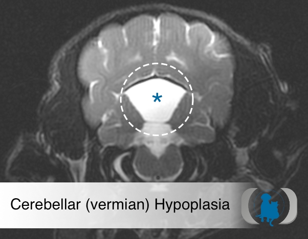
4) Vermian hypoplasia: Development of the cerebellar vermis is abnormal. When vermian hypoplasia is associated with cyst-like formation in the fourth ventricle, it is referred to as Dandy-Walker-like malformation in dogs. In most types of Dandy-Walker malformation, hydrocephalus is also present.

Pathophysiology

Various injuries to the brain have the potential to cause cerebellar hypoplasia. While trauma, hypoxia, toxins. and even genetic mutations have the potential to cause cerebellar hypoplasia, most cases are believed due to in utero exposure to Panleukopenia virus in the cat or Parvo virus in the dog.

Clinical Signs

Signs are noticeable when the affected animal first starts to walk, at around 2-3 weeks of age. Signs may include ataxia, hypermetria, dysmetria, broad-based stance, swaying of the body, fine tremors, and intention tremors of the head.

Diagnostic Tests

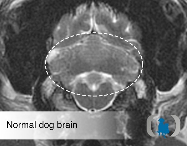
Advanced Imaging: Magnetic resonance imaging (MRI) of the brain shows a cerebellum that is significantly smaller than normal, with increased surrounding cerebrospinal fluid. When hypoplasia is mild, it can be challenging to distinguish congenital cerebellar hypoplasia from cerebellar degenerative diseases. With degenerative conditions, the cerebellum tends to be only slightly smaller or normal in size, with increased space between the cerebellar folia. Computed tomography of the brain has also been used to identify cerebellar malformations.

Therapy

No specific therapy is available. Symptomatic therapy which is largely environmental is generally beneficial. Limitation of the environment to include minimal opportunities for climbing and hence falling as well as the use of textured floor surfaces like carpeting or throw rugs/mats. Part compensation largely occurs due to learning of ways to overcome the inherent limitations that the condition presents.

Image Gallery

Midline sagittal T2 weighted MRI image of a normal dog brain
Midline sagittal T2 weighted MRI image of a normal dog brain. Notice the appearance of the normal cerebellum (dashed white circle) with its fine branching pattern of white matter tracts surrounded by a thin layer of cortical tissue.
Midline sagittal T2 weighted MRI image the brain of a juvenile chihuahua dog with cerebellar hypoplasia.
Midline sagittal T2 weighted MRI image the brain of a juvenile chihuahua dog with cerebellar hypoplasia. Notice the reduced volume of the cerebellum (dashed white circle) with an increased volume of CSF (blue asterisk) filling the space around the small cerebellum.
 Midline sagittal T2 weighted MRI image of the brain of a juvenile Maltese dog with hypoplasia of the cerebellar vermis
Midline sagittal T2 weighted MRI image of the brain of a juvenile Maltese dog with hypoplasia of the cerebellar vermis. Notice the absence of the cerebellar vermis (arrows) with an increased volume of CSF (blue asterisk) filling the space normally filled by the vermis.
Midline sagittal T2 weighted MRI image of the brain of a juvenile mixed breed  dog with cerebellar aplasia.
Midline sagittal T2 weighted MRI image of the brain of a juvenile mixed breed dog with cerebellar aplasia. Notice the absence of the entire cerebellum with an increased volume of CSF (blue asterisk) filling the space normally filled by the cerebellum.
 Axial T2 weighted MRI image of a normal dog brain at the level of the caudal fossa
Axial T2 weighted MRI image of a normal dog brain at the level of the caudal fossa. Notice the appearance of the normal cerebellum (dashed white ellipse) with its fine branching pattern of white matter tracts surrounded by a thin layer of cortical tissue.
 Axial T2 weighted MRI image of the brain at the level of the caudal fossa of a juvenile chihuahua dog with cerebellar hypoplasia
Axial T2 weighted MRI image of the brain at the level of the caudal fossa of a juvenile chihuahua dog with cerebellar hypoplasia. Notice the reduced volume of the cerebellum (dashed white ellipse) with an increased volume of CSF filling the space around the small cerebellum.
Axial T2 weighted MRI image of the brain of a juvenile Maltese dog with hypoplasia of the cerebellar vermis at the level of the caudal fossa.
Axial T2 weighted MRI image of the brain of a juvenile Maltese dog with hypoplasia of the cerebellar vermis at the level of the caudal fossa. Notice the absence of the cerebellar vermis (dashed white elipse) with an increased volume of CSF (blue asterisk) filling the space normally filled by the vermis.
Axial T2 weighted MRI image of the brain of a juvenile mixed breed  dog with cerebellar aplasia at the level of the caudal fossa
Axial T2 weighted MRI image of the brain of a juvenile mixed breed dog with cerebellar aplasia at the level of the caudal fossa. Notice the absence of the entire cerebellum with an increased volume of CSF (blue asterisk) filling the space normally filled by the cerebellum.