In 2003 AVMI became the first veterinary practice in California to install a multislice spiral CT scanner dedicated to small animal patient care. With the installation of our new 80 slice spiral CT, AVMI once again demonstrates our dedication to providing the best service for our patients and referring veterinarians.The massive leap forward in technology that this new state-of-the-art CT scanner offers allows us to break new ground in numerous aspects of veterinary diagnostic imaging:
No More Anesthesia
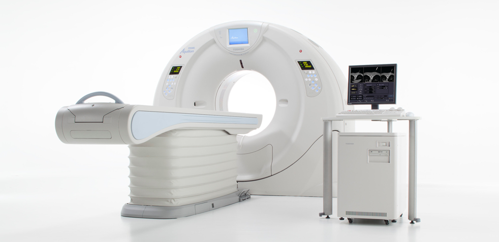
The ability to collect 80 slices per gantry rotation along with the industry leading 0.350 second gantry rotation time offered by our new Toshiba PRIME 80 slice CT scanner allows us to offer advanced diagnostic imaging of feline and small canine patient's without the need for chemical restraint. The ability to image a large dog's entire anatomy in a matter of seconds allows us to perform advanced diagnostic imaging procedures for even large patient's without the need for general anesthesia.
Perfusion (4D CT) imaging of endocrine tumors
The ability to image 4 cm of contiguous patient anatomy close to 3 times per second allows us to perform real-time perfusion imaging. This technique sometimes called 4D CT has been shown to be very useful in diagnosing numerous endocrine neoplasias.
Parathyroid adenoma:
Currently the diagnosis of small eutopic parathyroid adenomas and ectopic parathyroid adenomas of any size is extremely challenging in veterinary medicine. Because parathyroid scintigraphy using 99mTc-Sestamibi, which is very useful in people, has been shown to be of minimal utility in small animals, high resolution ultrasound is currently the initial diagnostic imaging modality of choice in dogs suspected of hyperparathyroidism secondary to a parathyroid adenoma. The increased temporal resolution provided by current CT scanners able to perform real-time perfusion (4D) CT is likely to make this modality the new diagnostic imaging modality of choice for diagnosing dogs with parathyroid adenomas in either eutopic or ectopic locations.
Insulinoma:
Currently the diagnosis of insulinoma in dogs is very challenging with arterial phase CT imaging already currently considered the gold standard diagnostic tool. Previous reports have been limited to devices capable of acquiring < 16 slices per gantry rotation. Our new 80 slice slice scanner should make the routine diagnosis of insulinoma comparitively much more straightforward and routine.
CT Fluoroscopy
SUREFluoro-CT Fluoroscopy permits real-time image reconstruction to display 3 simultaneous images obtained by combining data from the area detector. Moreover, SUREFluoro employs:
- Volume ONE shot, which is CT fluoroscopy volumetric scanning with MPR oblique display.
- MPR and oblique image guidance ensures accurate needle positioning with complete confidence during complex biopsy procedures, saving time and improving patient safety.
- Half scan can be selected for the scan mode, and the exposure angle can be specified.
- Single image viewing rate is 12 fps in a 512 matrix.
- All operations are performed at tableside. The operator is able to control table movement, gantry movement, and X-ray exposure while observing the progress of the procedure.
- Tilting can be performed from the extension operating panel.
- The last-image hold feature maintains the latest image while the beam is switched off.
Metal Artifact Reduction
SEMAR (Single Energy Metal Artifact Reduction) - utilizes a sophisticated reconstruction algorithm to reduce artifacts caused by metal while improving visualization of the implant, supporting bone and adjacent soft tissues for an accurate diagnosis. This new software feature will allow accurate CT evaluations in areas previously compromised by the potentially marked beam hardening artifacts that surround metallic implants including triple pelvic osteotomy bone plates and hip prostheses.
Adaptive Iterative Dose Reduction
AIDR 3D is the third generation in the evolution of Toshiba’s iterative reconstruction technology. AIDR 3D is an iterative algorithm intended to reduce pixel noise from the original data, the results analyzed, and the process repeated until the target level of noise-reduction is achieved. This iterative algorithm is superior in reducing background noise while preserving diagnostic information compared to non-iterative approaches. AIDR 3D can be integrated into all acquisition modes for routine clinical use and is able to reduce pixel noise magnitude in a way that allows radiation dose reduction. This feature will allow us to achieve diagnostic results while limiting the radiation dose to the patient.